

Research
My research background, vision and future interests

Research Background
My academic journey and research training have consistently centered on uncovering the molecular mechanisms by which RNA-protein interactions and phase-separated RNP condensates regulate gene expression, RNA metabolism, and stress responses in both bacterial and eukaryotic systems. I earned my Ph.D. in Biochemistry with Dr. Chakrabarti from Freie Universität Berlin, Germany, where I investigated the role of RNA helicase UPF1 in human histone mRNA decay. This work provided key insights into RNA surveillance and decay mechanisms in eukaryotes. Prior to my Ph.D., I worked at Heidelberg University in Germany and Indian Institute of Technology Guwahati on proteostasis and plant proteases, gaining diverse molecular biology skills. My first postdoctoral training at Wayne State University with Dr. Schrader has been instrumental in establishing my niche in RNP condensate biology where I led the first proteomic study of bacterial biomolecular condensates (BR-bodies), and contributed to multiple collaborative projects examining the dynamics, composition, and function of RNA decay condensates. These studies demonstrated how bacterial RNA binding proteins forms phase-separated condensates that regulate mRNA decay and enzymatic activity. Now, at the Fox Chase Cancer Center with Dr. Chen, I am building on this foundation to investigate how nuclear biomolecular condensates undergo structural and functional remodeling in response to stress and how their dysregulation contributes to cancer pathogenesis.
Research Vision
Proteins and RNAs do not function solely as individual molecular entities, they also exhibit emergent mesoscale behavior by self-organizing into dynamic biomolecular assemblies. These mesoscale structures possess physical and biochemical properties that are fundamentally distinct from those of their individual components, enabling new mechanisms of cellular regulation. By concentrating enzymes and substrates, selectively partitioning biomolecules, buffering molecular fluctuations, and creating specialized reaction environments, these assemblies can enhance biochemical reactions, spatially organize molecular processes, sequester regulatory factors, and give rise to functions that cannot be achieved by isolated proteins or RNAs. Understanding how mesoscale organization emerges and how it governs cellular function is a fundamental challenge in modern cell biology. Biomolecular condensation by phase separation is the prominent example of such mesoscale behavior which underlies the formation of Membraneless organelles or Biomolecular condensates in a cell. These condensates are widespread and play essential roles in organizing key RNA related processes including transcription, pre-mRNA splicing, RNA processing, localization and degradation and their aberration is increasingly linked to cellular malfunction and disease states. My research program is driven by a central vision - Biomolecular condensates act as programmable regulatory hubs that control RNA fate, and their dysregulation represents both a fundamental mechanism of disease and an untapped therapeutic opportunity. To address this, my work integrates biochemistry, cell biology, and advanced imaging approaches including optogenetics, two-photon microscopy, and 3D holotomography to define the biochemical and biophysical principles governing RNA-associated condensates across both bacterial and human systems.
Interests & Future Work
01
Investigating biomolecular condensate Assembly, maintenance, and disassembly
Biomolecular condensates are dynamic, membrane-less compartments that organize essential biochemical processes within cells. Although their overall formation in live cells takes minutes to hours, the key molecular events such as nucleation, client recruitment, and disassembly occur much faster, on timescales ranging from nanoseconds to seconds. Understanding these rapid spatiotemporal dynamics is crucial for explaining how condensates form, function, and respond to changes, and for developing strategies to modulate their behavior in disease.
However, capturing and controlling these transient events in living cells remains challenging, highlighting the need for precise, real-time experimental approaches. To address this, my work combines quantitative live-cell microscopy with in-vitro reconstitution. Live-cell imaging allows direct observation of condensate behavior in its native cellular context, while test-tube reconstitution provides a controlled system to study condensates and their components in isolation. Together, these complementary approaches enable a more complete and mechanistic understanding of condensate biology.

Live-cell imaging set up
Test-tube reconstitution of a minimal biomolecular condensate: Recombinantly purified bacterial Ribonuclease E and purified RNA was used to form Ribonuclease E condensate. This setup was used to test the recruitment of various client proteins.


Live-cell optogenetic tool to investigate condensate biogenesis: Here a nuclear condensate forming protein is fused with light responsive protein and mCherry, and the client protein is tagged with eGFP. When illuminated with 488 nm laser, the light responsive module brings the condensate forming molecules to form a condensate. This condensate in turn recruits the client protein. This method allows to determine the recruitment kinetics.
02
How do multi-pathway proteins achieve specificity?
A fundamental challenge in biology is understanding how multifunctional molecules achieve specificity within complex cellular networks. Many proteins participate in multiple biological pathways by interacting with different molecular partners and acting on distinct substrates. For example, protein X functions in RNA processing, RNA turnover, and the assembly of larger molecular complexes. Despite their involvement in diverse cellular processes, these proteins execute highly specific functions in a context-dependent manner. What mechanisms enable such multifunctional proteins to distinguish among competing pathways and selectively regulate distinct biological outcomes?
My research addresses this question by investigating the mesoscale organization of ribonucleoprotein (RNP) assemblies and biomolecular condensates. These dynamic, membrane-less structures organize proteins and nucleic acids into specialized biochemical environments that can influence molecular interactions and cellular behavior. Using an interdisciplinary approach that combines biochemical reconstitution, quantitative imaging, molecular genetics, and cell biology, I examine how condensates assemble, how they selectively recruit proteins and RNAs, and how their physical properties shape biological function. By studying these organizational principles across diverse biological systems, my work seeks to uncover general mechanisms through which cells compartmentalize biochemical activities and achieve functional specificity. Understanding how condensates organize multifunctional proteins into distinct molecular environments may provide a mechanistic framework for explaining specificity within complex cellular networks.

03
Reprogramming Nuclear Condensates in Cancer: Mechanisms, Dysfunction, and Therapeutic Control
Eukaryotic Nucleus is enriched with biomolecular condensates and they play essential roles in gene expression and regulation, including transcription, rRNA biogenesis, pre-mRNA splicing etc. Beyond their central functions in nuclear organization, they also act as sensors and regulators of cellular stress, responding to both specific and nonspecific perturbations such as anticancer drug treatment. These stress responses can drive the formation of aberrant condensates, which may disrupt normal biochemical processes, acquire novel functions, alter their material properties, or sequester therapeutic agents, thereby contributing to drug resistance.
My research aims to dissect the biochemical and biophysical mechanisms underlying condensate-mediated stress sensing and regulation by integrating optogenetics, live-cell imaging, fluorescence lifetime imaging microscopy (FLIM), and complementary biochemical approaches. By systematically characterizing the properties and functional consequences of aberrant condensates in nuclear bodies such as the nucleolus, Cajal bodies, nuclear speckles, and histone locus bodies, this work will determine whether chemotherapy-induced condensate alterations enhance therapeutic efficacy or promote unintended outcomes such as drug resistance.
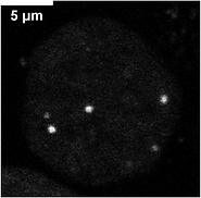

(Left) A normal spherical condensate in the nucleus exhibits a liquid-like nature. (Right) Upon treatment of the cells with an anti-cancer drug which is non-specific to this particular condensate, converts to irregular shape and exhibits gel-like nature.

Fluorescence Life-time
imaging (FLIM) can reveal material state of a condensate (whether a condensate is liquid-like or solid-like). When FLIM
is combined with FRET, it provides powerful approach to probe quantitative protein-protein interactions within a condensate